News
CSIC's Radiopharmacy Service Line now operational and available for research study support.
- CSIC's Radiopharmacy service line is now fully operational and producing radiopharmaceuticals in our new HSRB-II lab facility for use in human-subject research studies that utilize CSIC's PET/CT or PET/MR, imaging services, or for PET research scans completed at an EHC PET scanner. Investigators can contact Ron Crowe, RPh, BCNP, (Radiopharmacy Manager) or Steven Liang, PhD (CSIC PET/Radiopharmacy Program Director) to request information about radiopharmacy services, or see our Radiopharmacy Services resource page.
11.7T Animal MRI service line (HSRB-II) now operational and available for research study support.
- Contact Shella Keilholz, PhD (CSIC Animal MRI Program Director) or Jaekeun Park, PhD (CSIC Animal MRI Scientist) to discuss protocol imaging support needs and technical capabilities of CSIC's newest animal MRI scanner.
HSRB II Facility - CSIC Location
Emory's Center for Systems Imaging Core Now Located in State-of-the-Art Facility in the HSRB II
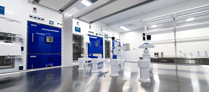
The Center for Systems Imaging Core is now located in a state-of-the-art facility in Emory's newest 350,000 square foot specialized research facility, Health Sciences Research Building II (HRSB II). The 17,800 square foot facility contains a 3.0 Telsa human MRI scanner, a 7.0 Tesla MRI scanner (the first in Georgia), a human PET/CT system, as well as study participant waiting area, consent rooms, changing rooms, and PET support/recovery rooms. The center also has a new 16.5 MeV proton dual target medical cyclotron and 10 hot cells that support new radiopharmacy and radiochemistry facilities. The pre-clinical section of the center includes an 11.7 Telsa MRI scanner and a micro-PET/CT scanner. Space has also been reserved for expansion of future core imaging equipment and support space.
This new equipment and space enables Emory investigators to use imaging to see into the body in ways that have not been done before. These capabilities give investigators tools they can use to develop new diagnostic markers, understand health and disease, and search for new treatments.
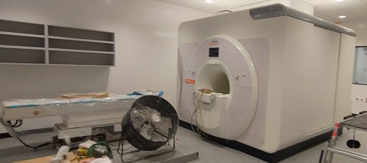
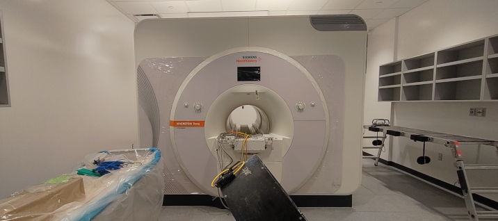
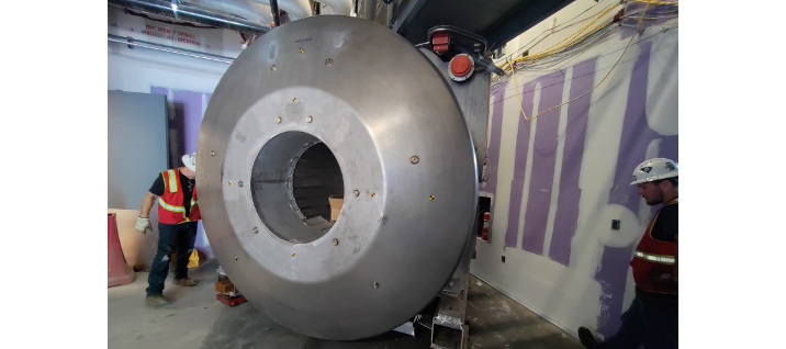
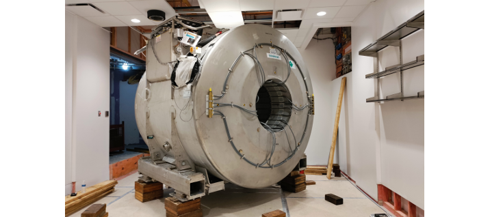
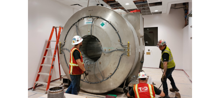
- Siemens Terra 7T Ready for Calibration
Siemens Terra 7T Mechanical/Electronic Installation Accomplished
While the electro-magenetic field shielding for the 7T scanner room is closed, Siemens accomplished the mechanical and electronic installing of the system. It is now ready to be calibrated. We are expecting a working 7T system in about a month.
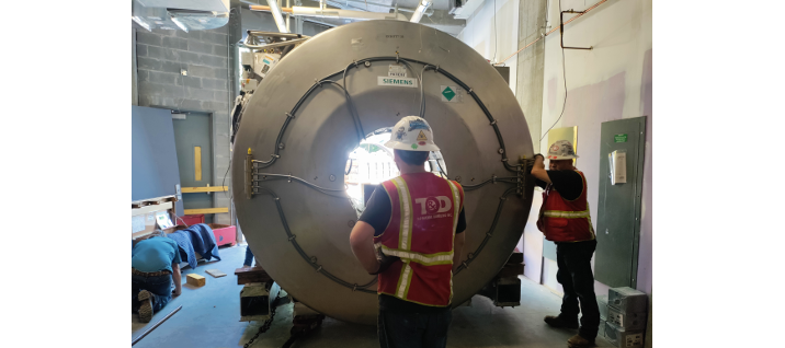
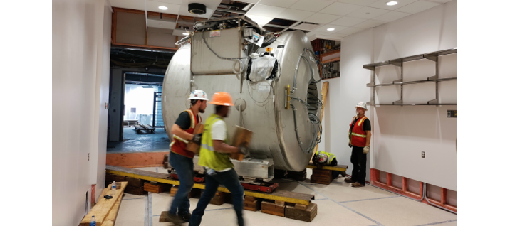
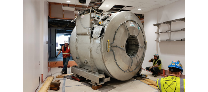
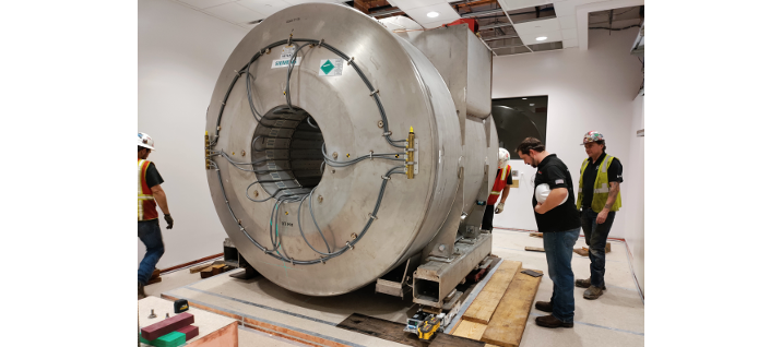
- Siemens Terra 7T MRI arrived HSRB II
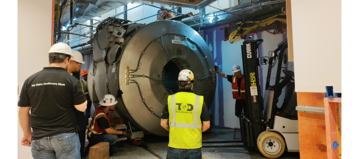
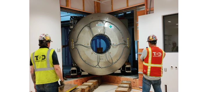
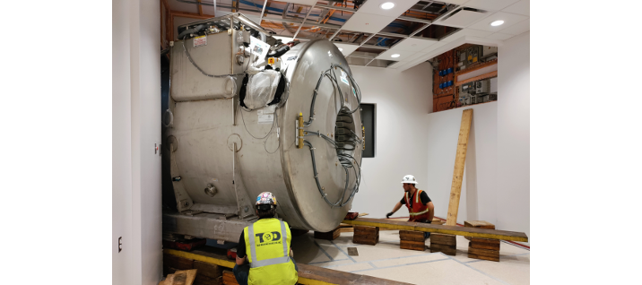
Siemens Terra 7T moving in
On May 14th, 2022, we are excited watching the Siemens 7T Terra MRI scanner moving into the designated suite in the under construction HSRB II building.
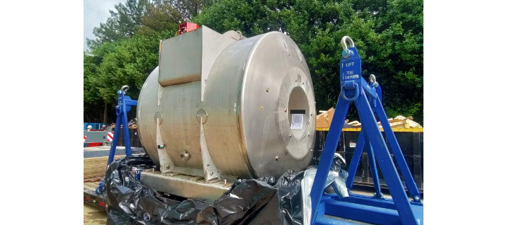
09:55 - T1 magnet arrived
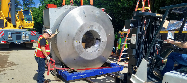
10:47 - Magnet off the truck. Loading frame being removed.
11:45 - Magnet moving to the door
11:51 - View from inside building - Some overhead structures have to be removed to yield enough clearance for the magnet
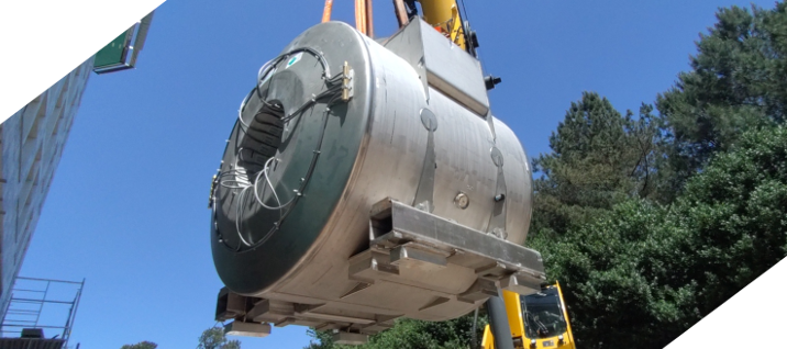
13:06 - Magnet in HSRB II building
13:14 - From entrance hall to just fit corridor - only 3 inches each side to the wall and over head structures
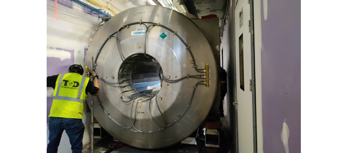
13:22 - Going through the corridor
13:24 - Approaching the scanner room
13:27 - View from inside scanner room
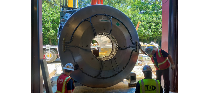
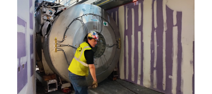
13:45 - Building bridge to roll the magnet in
14:17 - Magnet on the bridge
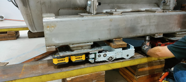
14:29 - Details of the strong tiny robot that drives the magnet
14:36 - Magnet fully indoor
14:52 - Bridge removed, magnet ready to be lowered down to the concrete seat
15:42 - Magnet down to the ground level, ready to be turned to the designed orientation
16:02 - Magnet reached final location
16:39 - All shipping frames removed. Magnet on rubber feet at designated location. Job well done!
- CSIC's new home - HSRB Phase II
- The Emory Brain Imaging Project
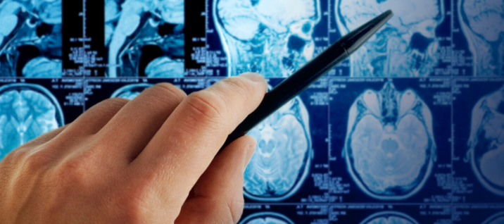 The Emory Brain Imaging Project is one facet of the Emory Healthy Aging Study, the largest clinical research project that has ever been conducted by Emory University. In totality, the study focuses on enhancing our knowledge of the aging process and addressing the growing number of people who are being diagnosed with age-related diseases such as Alzheimer’s Disease. Participants in this study undergo a series of tests every two years, which involves collecting blood samples, retinal imaging, cognitive testing, gait analysis, cerebrospinal fluid analysis, vascular assessment and most importantly, Magnetic Resonance Imaging (MRI) of the brain, conducted by the Emory Brain Imaging Project. The MRI imaging is a critical component to this study since this modality has the unique capability of collecting structural and functional data from the brain that will help researchers identify biological markers that are predictive of common, age-related diseases. Ultimately, this study will change the way we understand the aging process and support the development of treatments for Alzheimer’s Disease and other common age-related diseases.