About
Located at two sites at Emory University’s main campus, the Robert P. Apkarian Integrated Electron Microscopy Core (IEMC) provides services and training on conventional transmission electron microscopy (TEM), cryo-transmission electron microscopy (Cryo-TEM), single particle cryo-electron microscopy (SP-Cryo-EM), cryo-electron tomography (Cryo-ET), conventional scanning electron microscopy (SEM), and cryo-high resolution scanning electron microscopy (Cryo-HRSEM). The IEMC supports academic, clinical, and industry users. Its focus and technical expertise aim to generate structural data from biological and non-biological specimens to support research in basic, biomedical, and material sciences and engineering.
Robert P. Apkarian, Ph.D.
Our facility is named in honor of Dr. Robert P. Apkarian, an internationally recognized microscopist, who developed chromium-coating methodologies and who was a pioneer of the field of cryo-scanning electron microscopy. He pursued the scientific frontiers of the micro-world, and combined his love of the natural sciences with his passion for electron microscopy.
Core Leadership
Core Team
Sondip K. Biswas, Ph.D.
Associate Scientist
Office: Room E106. Cherry Logan Emerson Hall
Phone: 404-712-8674
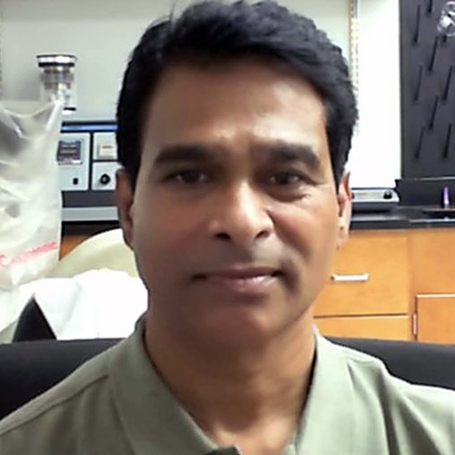
Sondip provides technical expertise and support for SEM and TEM imaging projects across a variety of sample types and project workflows. Over his many years of experience, his research has focused on structural biology and ophthalmic science, using advanced electron microscopy to investigate mitochondrial ultrastructure and cell-to-cell junctions in the eye. Sondip frequently collaborates with Georgia Research Alliance investigators, contributing to interdisciplinary biomedical research. He has published extensively on cholesterol distribution and the formation of aquaporin and gap junctions in lens fiber cells, findings that provide important insight into the mechanisms underlying cataract formation. At the IEMC, he supports users through complete workflows on TEM and SEM projects, from experimental design to data acquisition, and analysis, making sure that investigators obtain high-quality structural data for their publications.
Jack Botting, M.Sc.
Assistant Academic Research Scientist
Office: Room E106. Cherry Logan Emerson Hall
Phone: 404-712-8674
Jack is an expert in cryo-FIB-SEM and cryo-EM, with special emphasis and user training expertise in lamella milling preparation for cryo-ET applications. He supports cryo- and room temperature FIB-SEM projects including volume EM and lamella milling. He also carries out FIB-SEM volume and cryo-ET data analysis using semiautomated software and machine learning and has years of experience in high-throughput tilt series acquisition and processing. This includes software for visualization, segmentation, and sub-tomogram averaging. Jack also contributes with his expertise to cryo-EM sample preparation, including plunge freezing and optimization of grid and sample conditions for FIB-SEM and cryo-ET.

Srihari (Ravi) Koripella, Ph.D.
Scientist
Office: Room G236. Biochemistry Connector. Rollins Research Center
Phone: 404-727-8960
Ravi is a structural biologist with a Ph.D. in Cell and Molecular Biology. He oversees cryo-TEM workflows at the Biochemistry connector site, managing the day-to-day operation of ThermoFisher Scientific Talos L120C and Talos Arctica transmission electron microscopes, as well as plunge freezing and grid-optimization equipment including the Vitrobot and Gatan CP3. Ravi supports data acquisition and provides hands-on training to new users in cryo-TEM sample preparation and microscope operation and assists researchers with data processing using software on the IEMC’s high-performance computing workstations. With more than ten years of experience supervising students and teaching laboratory courses in molecular biology, biotechnology, and cell biology, he brings strong expertise in cryo-sample preparation, grid optimization, and experimental design, and is a valuable resource for investigators pursuing 2D cryo-TEM, single particle cryo-TEM, and cryo-electron tomography applications.
ORCiD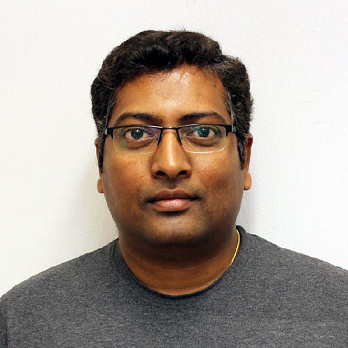
Jiawei Li, Ph.D.
Assistant Academic Research Scientist
Office: Room E106. Cherry Logan Emerson Hall
Phone: 404-712-8674
Jiawei is a structural biologist with a Ph.D. in molecular miophysics and extensive hands-on expertise in transmission electron microscopy techniques including room temperature and cryo-TEM sample preparation methods. She joined the IEMC after years of experience supporting biomedical research through advanced electron microscopy workflows in a user-focused facility where she contributed to training and mentoring, data acquisition, data processing and analysis. Jiawei will support room temperature structural analysis of tissue and cell cultures, applying TEM single imaging and tomography acquisition methods. Her background in specialized cryo-TEM applications also supports the consolidation of cryo-workflows including cryo-electron microscopy of vitrified sections (CEMOVIS) and cryo-immunolabeling techniques.

Casey F. Rowe, Ph.D.
Assistant Academic Research Scientist
Office: Room E106. Cherry Logan Emerson Hall
Phone: 404-712-8674
Casey is an experienced electron microscopist with extensive usage of advanced systems for TEM, STEM and SEM workflows, including FIB-SEM lamellae preparation. His experience in materials science and characterization of 2D materials and crystals is essential for the optimization of TEM, SEM, micro-ED workflows, user training, and the establishment of SOPs for various EM applications. Casey also has experience in EM data processing using MATLAB and Python and assists users during data processing. He has several years of experience supervising students on sample preparation and data acquisition for electron microscopy and FIB milling procedures, an important resource to support, and train new investigators.

Ted J. Whitworth, Ph.D.
Associate Scientist
Office: Room E107. Cherry Logan Emerson Hall
Phone: 404-712-8491
Ted is an expert structural biologist with a Ph.D. in Experimental Pathology and more than 25 years of experience in ultrastructural pathology and electron microscopy. With extensive experience in operating and maintaining TEM instrumentation and preparing diverse specimens (microorganisms, viruses, human tissues, and cell cultures) for structural biology applications, Ted is highly skilled in negative staining, immunogold labeling, plastic embedding, thin and ultrathin sectioning, high-pressure freezing, freeze substitution, and Tokuyasu ultrathin cryo-sectioning. He has taught courses in biology, microbiology, ultrastructure, anatomy, and physiology and has trained numerous investigators in electron microscopy workflows. At the IEMC, he supports users through experimental design, sample preparation, data acquisition, and analysis, helping researchers generate high-quality, publication-ready structural data.