Services
The IEMC provides services and expertise in all aspects of transmission and scanning electron microscopy including conventional and cryo preparation of cells and tissues, negative staining, metal and protein shadowing, and freeze-fracture.
Our services include room temperature and cryogenic Transmission and Scanning Electron Microscopy imaging and sample preparation for biological and materials science specimens, energy-dispersive Spectroscopy (EDS) for elemental analysis, room temperature and cryogenic ultramicrotomy, high-pressure freezing (HPF) and self-pressurized rapid freezing (SPRF), cryo/freeze-substitution, ultrathin metal film coating, and immunocytochemistry.
Our experienced staff is available to help our users with basic image analisis and advanced image processing workflows for grants and publications.
Transmission Electron Microscopy
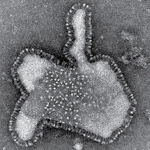
The IEMC houses five transmission electron microscopes suitable for imaging biological and non-biological specimens prepared using a number of methods both at room temperature and under cryo conditions. Image: RSV particle from the IEMC image library.
Cryo-Transmission Electron Microscopy
Four of our TEMs are equipped with cryo-capabilities using Gatan 626, 714, or ELSA cryo-holders for single-tilt and high-tilt imaging of biological specimens or materials. Our Talos Arctica comes with an autoloader system that allows the loading of 12 cryo-EM samples for fast grid mapping and high-throughput image acquisition of high-resolution, single-particle data. Image acquisition is done on a Gatan K3 direct electron detector (Talos Arctica), or a Direct Electron DE20 detector (JEOL JEM-2200FS). Image: Measles virus particle from the IEMC image library.
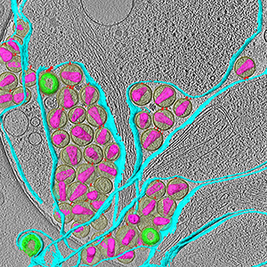
Cryo-Transmission Electron Tomography
Our 200 kV, JEOL JEM-2200 FS, with an in-column Omega Filter, a Gatan 714 high-tilt cryo-holder and a Direct Electron detector (DE20) is capable of collecting high quality tomography data. Image: Segmented tomogram of HIV-1 virions attached to plasma membrane (DOI: 10.1128/JVI.01880-15).
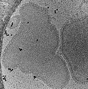
Scanning Electron Microscopy with Energy Dispersive Spectroscopy
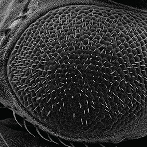
We have a Field Emission Scanning Electron Microscope (SEM, JEOL JSM-IT700HR), acquired with funds from the Georgia Research Alliance. This instrument is fitted with secondary and back-scattered electron detection and it is capable of low and high-vacuum SEM. It has a JEOL JED-2300 Dry SDD, fully-embedded, energy-dispersive microanalysis system to carry out Energy-Dispersive Spectroscopy (EDS) for elemental analysis. Image: Fly eye from the IEMC image library.
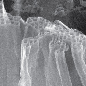
Plasma Focused Ion Beam-Scanning Electron Microscopy
Funded through an NIH High-End Instrumentation Grant, Emory's School of Medicine, Emory College of Arts and Sciences, and the Department of Chemistry, a state-of-the-art Focused Ion Beam - Scanning Electron Microscope (ThermoFisher Hydra Bio Plasma FIB-SEM) has been installed at our Emerson site. This state-of-the-art instrument is capable of cryo and room temperature applications and has four switchable ion species: Xe, Ar, O, and N. Samples can be prepared by plunge-freezing in liquid ethane, high-pressure freezing, and metal coating. An integrated Fluorescent Light Microscope (iFLM, Delmic Meteor) system allows Emory researchers to perform Correlated Light and Electron Microscopy (CLEM) without the need for sample exchange between instruments. Image: Cryo-SEM of amyloid-beta oligopeptides self-assembled into nanotubes (DOI: https://doi.org/10.1039/B701029J).