News
News from IEMC
Electron micrographs used for these figures were acquired at the IEMC. Thank you for acknowledging our contribution.
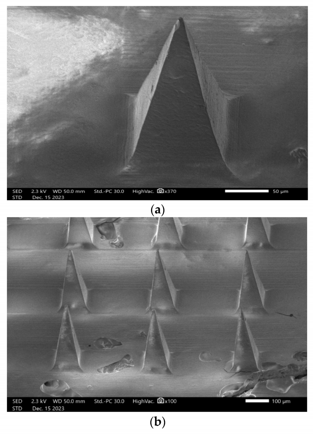
This publication in the Journal Pharmaceutics describes the development and evaluation of a transdermal delivery system (microneedles) for the schizophrenia medication lurasidone. Through the Georgia Research Alliance’s Core Facilities Partnership, Dr. Ajay K. Banga's group at Mercer University’s Center for Drug Delivery Research utilized Emory University’s state-of-the-art JEOL JSM-IT700HR scanning electron microscope to study the characteristics of these microneedles. At the Robert P. Apkarian Integrated Electron Microscopy Core, we take pride in contributing to transformative projects by Georgia researchers. Find the article here: https://www.mdpi.com/1999-4923/16/3/308
Radmard A, Banga AK. Microneedle-Assisted Transdermal Delivery of Lurasidone Nanoparticles. Pharmaceutics. 2024 Feb 22;16(3):308. doi: 10.3390/pharmaceutics16030308. PMID: 38543202; PMCID: PMC10974263.
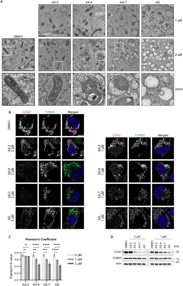
In this recent publication, researchers at Emory University’s Department of Hematology and Medical Oncology and the Winship Cancer Institute of Emory University describe the development and assessment of a class of bisbiguanides (AX-4 and AX-7, analogs of alexidine) as disrupters of mitochondria and cell invasion, adding them to the list of promising compounds aimed at limiting cancer metastasis. Utilizing transmission electron microscopy (TEM) methods and making use of our Georgia Research Alliance-funded Hitachi HT7700 (Hitachi Electron Microscope) and our JEOL JEM 1400 (JEOL USA) TEMs at the IEMC, the authors examined the effects of analog treatment on mitochondria ultrastructure. Find the article here: https://doi.org/10.1016/j.isci.2024.109591
Knippler, CM, Arnst, JL, Robinson, IE, Matsuk, V, Khatib, TO, Harvey, RD, Shanmugam, M, Mouw, JK, Fu, H, Ganesh, T, Marcus, AI, Bisbiguanide analogs induce mitochondrial stress to inhibit lung cancer cell invasion, ISCIENCE (2024), doi: https://doi.org/10.1016/j.isci.2024.109591.
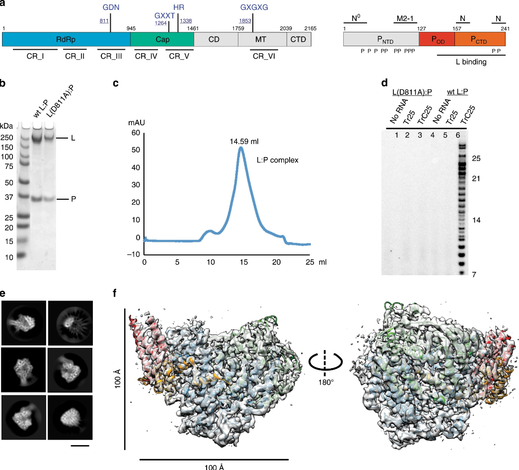
a - Schematic domain representation of the human respiratory syncytial virus (RSV) RNA polymerase (L:P complex), with labeled domain boundary (Alignment details in Supplementary Fig. 4). The domains with missing density are colored in gray. b - The SDS-PAGE gel shows the quality of the wild-type (wt) and mutant RSV RNA polymerase, wt L:P, and L(D811A):P, respectively (repeated ≥ 5 times). c - Elution profile of the purified L:P complex on the Superose 6 Increase 10/300 GL size-exclusion column (repeated ≥ 5 times). d - The RNA dependent RNA polymerization assays show that the purified wt polymerase is active and can synthesis RNA with specific RNA templates (lane 6) compared with that of catalytically inactive polymerase L(D811A):P (lane 3). e - Representative 2D class averages from the 200 kV cryo-EM dataset, selected amongst 125 classes. Scale bar: 100 Å. f - The final cryo-EM density map with the model (colored as a) in two orientations.
Cao D, Gao Y, Roesler C, Rice S, D’Cunha P, Zhuang L, Slack J,Domke M, Antonova A, Romanelli S, Keating S, Forero G, Juneja P, Liang B. 2020. Cryo-EM structure of the respiratory syncytial virus RNA polymerase. Nature Communications. 11(1):368
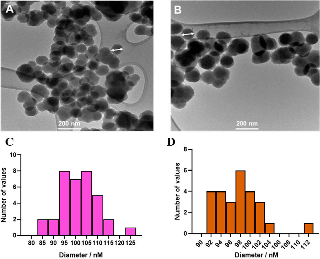
Teams from the Department of Biology and the Center for Diagnostics and Therapeutics at the Department of Chemistry at Georgia State University describe here the development of a sandwich immunoassay that combines silica nanoparticles and biorthogonal chemistries to enhance signal and lower the limits of detection of HIV-1 p24 antigen. Through the Georgia Research Alliance’s Core Facilities Partnership, dye-encapsulated, fluorescent silica nanoparticles were imaged by transmission electron microscopy at the IEMC, using our ThermoFisher Talos L120C, 120 kV, LaB6 TEM with a 4k × 4K Ceta CMOS detector (Thermo Fisher Scientific).
Jia T, Saikam V, Luo Y, Sheng X, Fang J, Kumar M, Iyer SS. Combining Bioorthogonal Chemistry with Fluorescent Silica Nanoparticles for the Ultrasensitive Detection of the HIV-1 p24 Antigen. ACS Omega. 2024 Mar 12;9(12):14604-14612. doi: 10.1021/acsomega.3c06136. PMID: 38559966; PMCID: PMC10976350.
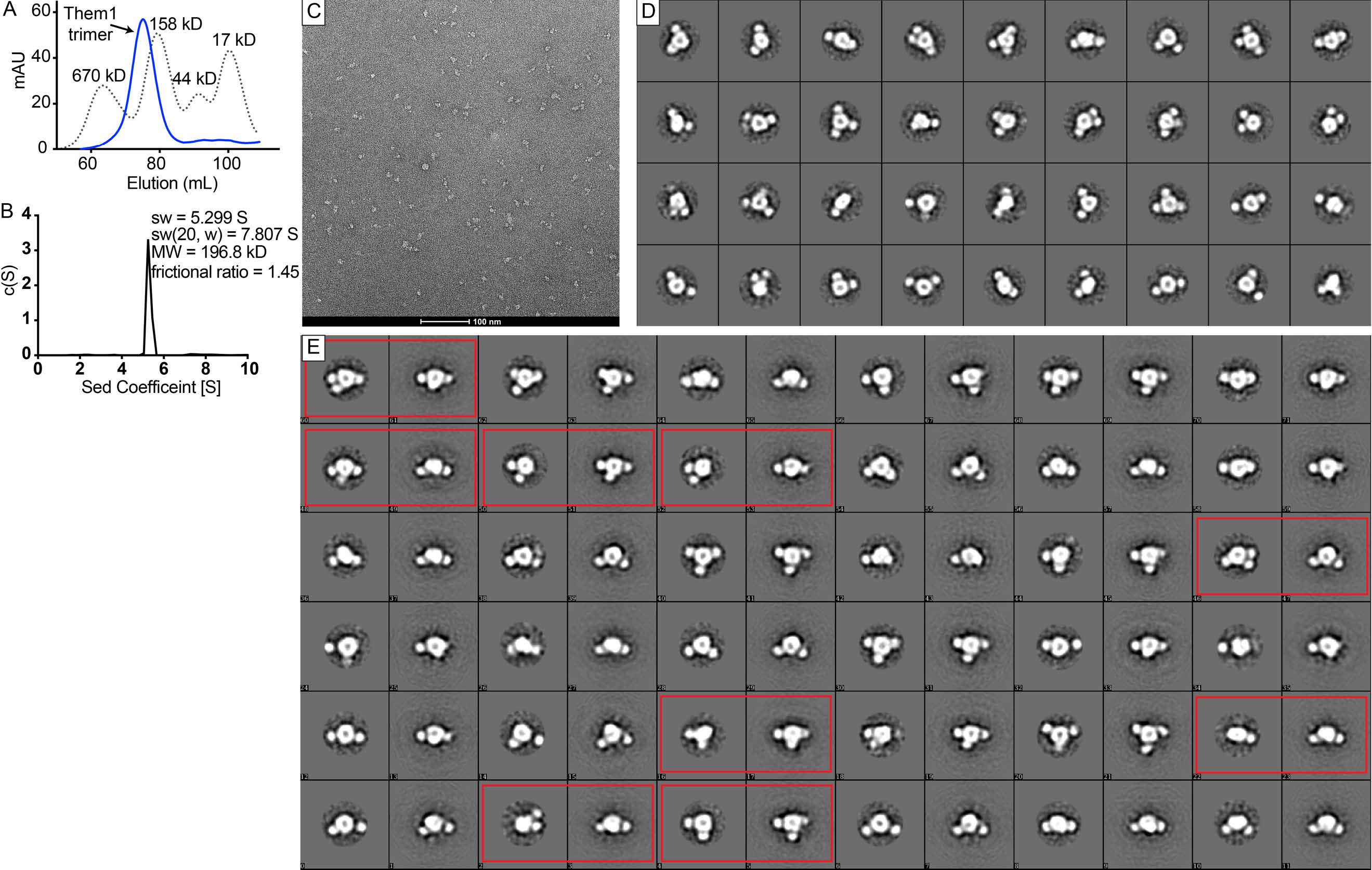
Negative stain single particle electron microscopy of Them1. A-B. DNterm-Them1 purifies as a homogenous trimeric complex (~196.5 kD) as determined by size exclusion chromatography (A) and analytical ultracentrifugation (B). A. Size exclusion chromatography of DNterm-Them1 (blue) and standards (dotted line). B. c(s) distribution from sedimentation velocity analytical ultracentrifugation of DNterm-Them1 complex. C. One image of DNterm-Them1 stained with uranyl formate spread across carbon coated Cu mesh grid collected on Talos 120 C Microscope at a magnification of 96,000X. D. 2D Class averages of Them1 using Relion 3.0. E. Comparison of 2D class averages (left) and reprojections (right) of 3D model generated with C3 symmetry to match the class average. Red rectangles highlight when reprojections of 3D model do not match class averages.
Tillman MC, Imai N, Li Y, Khadka, M, Okafor CD, Juneja P, Adhiyaman A, Hagen SJ, Cohen DE, Ortlund EA. 2020. Allosteric regulation of thioesterase superfamily member 1 by lipid sensor domain binding fatty acids and lysophosphatidylcholine. Proceedings of the National Academy of Sciences USA. Aug 20;202003877
IEMC News
Our ThermoFisher (TFS) Hydra Bio Plasma Focused Ion Beam – Scanning Electron Microscope (pFIB-SEM) is ready and available to researchers. The pFIB-SEM technology allows biomedical investigators in human health and human diseases to understand the three-dimensional organization of cellular environments and have access to state-of-the-art research techniques. This instrument is also equipped with a Delmic Meteor integrated Fluorescent Light Microscope (iFLM), which allows for Correlated Light and Electron Microscopy (CLEM) methods.
With this new technology, the IEMC will support structural research of cells and macromolecular complexes involved in infectious diseases, neurodegenerative disorders, heart and liver conditions, and cancer. Acquisition of this pFIB-SEM equipment with cryo-capabilities has immediate and profound impact by supporting a number of NIH-sponsored investigators at Emory University and members of the Georgia Core Facilities Partnership, who enjoy shared-access to each other’s state-of-the-art core facilities.
Electron Microscopy Events
Microscopy and Microanalysis - 2026
The meeting of the Microscopy Society of America will take place at the Baird Center in Milwaukee, WI from August 2-6, 2026. Link
SouthEastern Microscopy Society meeting - 2026
SEMS 61th annual meeting will be held May 11- 13, 2026 at the University of Georgia Center for Continuing Education and Hotel. Link