Resources
Our resources at IEMC are here to help you:
- Plan your project
- Write a grant or paper
- Learn about policies for accessing our microscopes
We are here to support your research. If you need assistance with anything not included here, simply email us at EMCore@Emory.edu.
Our range of electron microscopes and ancillary equipment includes:
IEMC Instrumentation - Cherry L. Emerson Site - Microscopes
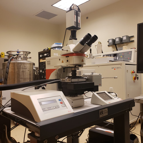
JEOL JEM-2200FS 200 kV TEM
Cherry Logan Emerson Hall. Suite E117
- The JEOL JEM-2200FS 200kV Field Emission TEM has two direct electron detectors, a Direct Electron DE-20 and a Gatan K2.
- Images can be acquired with improved contrast by the use of Zernike or hole-free phase plates.
- An in-column energy filter (Omega filter) is especially important for high-resolution cryo-electron tomography and cryo-electron microscopy of thick samples.
- SerialEM software is used for semi-automated acquisition of cryo-EM, cryo-ET, and microcrystal electron diffraction data.
- In addition to single-tilt, high-tilt, and dual-axis room temperature holders, two Gatan 914 cryo holders are available for tilt series acquisition and imaging of cryo-TEM specimens.
Please contact IEMC (iemc@emory.edu) to request training or use the link below to book this instrument.
Book this microscope

JEOL JEM-1400 120 kV TEM
Cherry Logan Emerson Hall. Suite E111
- The JEOL JEM-1400 120 kV LaB6TEM is equipped with a Gatan UltraScan, 2k x 2k, CCD camera.
- There are two Gatan 626 cryo holders available for acquisition of cryo-TEM data. Room temperature holders to carry out high-tilt applications and dual-axis tomography are also available.
- The JEOL JEM-1400 is capable of several modes of TEM, including electron tomography for room temperature and cryo applications.
- Semi-automated data acquisition using Serial EM is available for testing cryo-TEM grids, and a Minimum Dose System (MDS) function allows for imaging of beam-sensitive samples.
- The beam blocker allows for electron diffraction experiments.
Please contact IEMC (iemc@emory.edu) to request training or use the link below to book this instrument.
Book this microscope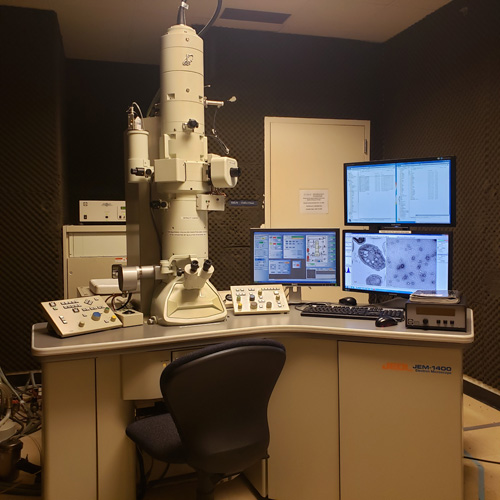
Hitachi HT7700 120 kV TEM
Cherry Logan Emerson Hall. Suite E116
- The Hitachi HT7700 TEM has a Tungsten filament, and it is used for imaging biological and materials samples with an AMT CCD camera.
- This instrument is capable of tilt imaging with a motorized goniometer which allows +/- 70° sample tilting.
Please contact IEMC (iemc@emory.edu) to request training or use the link below to book this instrument.
Book this microscope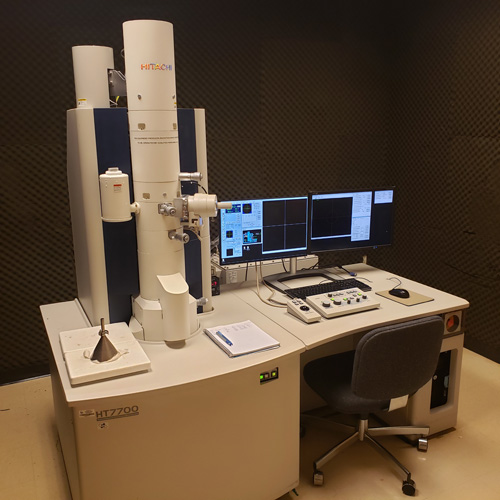
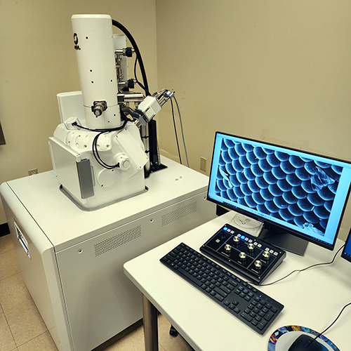
JEOL JSM-IT700HR SEM
Cherry Logan Emerson Hall. Suite E117
- The JEOL JSM-IT700HR is an analytical, high-resolution, field emission scanning electron microscope (SEM) capable of imaging under high and low vacuum conditions. With a large analytical chamber, this instrument is used for imaging biological samples and materials.
- A specialized MP-94370LSED Low Vacuum Secondary Electron Detector allows acquisition of structural data for soft, uncoated, hydrated samples.
- It is equipped with both a secondary electron detector and a high-sensitivity, multi-segment, solid-state backscattered electron detector.
- This instrument features a fully embedded JEOL JED-2300 energy-dispersive micro-analysis system (EDS) with a silicon drift detector (SDD) allowing elemental analysis of materials and biological samples.
Please contact IEMC (iemc@emory.edu) to request training or to schedule image acquisition on this instrument.
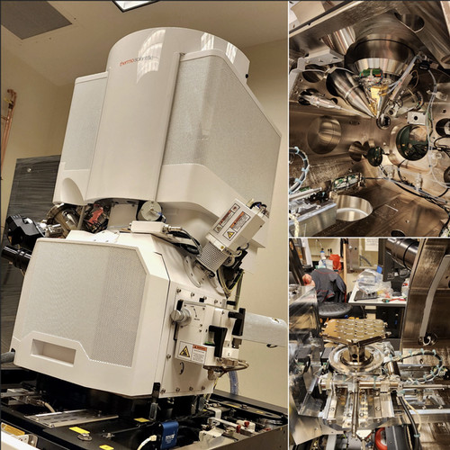
ThermoFisher Hydra Bio Plasma FIB-SEM
Cherry Logan Emerson Hall. Suite E115
- Thermo Scientific Hydra Bio Plasma Focused Ion Beam-Scanning Electron Microscope with a Delmic METEOR integrated Fluorescent Light Microscope (iFLM) for Correlated Light and Electron Microscopy (CLEM).
- This instrument is equipped with 4 ion sources (Xenon, Argon, Oxygen, and Nitrogen) to allow for multiple FIB-SEM applications on biological samples and materials.
- Spin-Mill capabilities increases throughput in room temperature volume EM by acquiring data in multiple regions of interest within a single sample.
- Cryo applications convert this instrument in a sample preparation tool for lamella milling, as well as a data acquisition tool for cryo volume EM.
Please contact IEMC (iemc@emory.edu) for more information.
Leica DM6 FS cryo-CLEM fluorescence microscope
Cherry Logan Emerson Hall. Suite E115
- The Leica Cryo-CLEM system for Correlated Light and Electron Microscopy (CLEM) is a cryo fluorescence microscope which allows the observation of frozen-hydrated samples from viruses to cells on cryo-electron microscopy (cryo-TEM) grids.
- Data acquisition on this instrument permits mapping of cryo-TEM grids and transferring of coordinates to a TEM for further, high-resolution, data collection with a semi-automated software.
Please contact IEMC (iemc@emory.edu) to request training or to schedule data acquisition on this instrument.
IEMC Instrumentation - Cherry L. Emerson Site - Sample Preparation Equipment
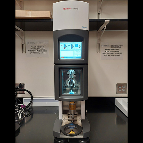
ThermoFisher Vitrobot Mark IV
Cherry Logan Emerson Hall. Suite 115
- The ThermoFisher Vitrobot Mark IV is used for plunge freezing aqueous solutions, cell suspensions, and on-grid grown cells.
- Vitrification can be done in liquid ethane, liquid propane, or an ethane/propane mix to prepare samples for cryo-TEM or cryo-ET data acquisition.
Please contact IEMC (iemc@emory.edu) to request training or use the link below to book this instrument.
Book this instrument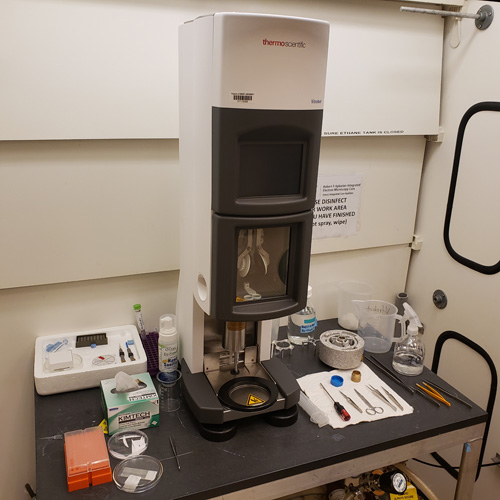
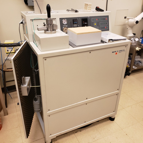
BAL-TEC HPM 010 High Pressure Freezing Machine
Cherry Logan Emerson Hall. Suite 115
- This high-pressure freezing machine is used to prepare frozen specimens, including thick samples, suspensions, and monolayer cell cultures, without the need of cryoprotectants.
- By measuring temperature and pressure in real time, this machine freezes samples with liquid nitrogen under high-pressure to avoid the buildup of ice crystals.
- High pressure frozen samples can be used for downstream electron microscopy applications or further processed by freeze substitution.
Please contact IEMC (iemc@emory.edu) to request training or to schedule sample preparation with this instrument.
Leica EM AFS2 Freeze Substitution Apparatus
Cherry Logan Emerson Hall. Suite 115
- This instrument allows for freeze substitution and progressive lowering of temperature (PLT) techniques.
- It creates the appropriate environment for low temperature embedding and polymerization of resins.
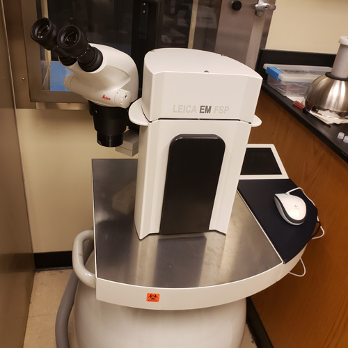
- The system is supported by a Leica EM FSP (freeze substitution processor), an automatic reagent handling system, which dispenses reagents for both freeze substitution and PLT applications.
- There is LED illumination from within the chamber and a stereomicroscope for viewing and positioning of samples.
Please contact IEMC (iemc@emory.edu) to request training or to schedule sample preparation with this instrument.
Microtomy Equipment
Cherry Logan Emerson Hall. Suites E109 and E115
- Several ultramicrotomes are available for processing plastic-embedded samples for TEM applications including a Leica UC6rt, a Leica Ultracut S, and an RMC PowerTome PT-XL ultramicrotome.
- A Leica UC6i/FC6 cryo-ultramicrotome is available to prepare biological specimens for cryo-electron microscopy of vitrified sections (CEMOVIS). In some cases, cryo-ultramicrotomy is used to process materials.
- A Leica Enuity ultramicrotome allows for semi-automated sectioning and process visualization with an integrated camera.
- Diamond knifes for room temperature and cryo applications are used for block trimming, as well as semithin, and ultrathin sectioning, depending on the application and sample.
Please contact IEMC (iemc@emory.edu) to schedule sample preparation with these instruments.

SEM Sample Preparation Equipment
Cherry Logan Emerson Hall. Suites 110 and 112
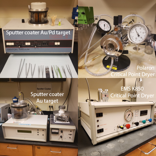
- Several instruments are available for preparing samples for SEM applications including two sputter coaters for gold or gold/palladium coating and two critical point dryers (CPDs) to prepare biological samples and soft materials for SEM.
- The EMSCOPE SC-500 Magnetron Sputter system can deposit thin layers of gold onto various types of samples for SEM.
- The Denton Desk II sputter coater has a gold/palladium target for the preparation of SEM samples.
- The Polaron CPD can hold large samples or can deposit many small samples for high-throughput preparation.
- The EMS K850 CPD can prepare samples using a variety of holder types including microscope slides.
Please contact IEMC (iemc@emory.edu) to schedule sample preparation with these instruments.
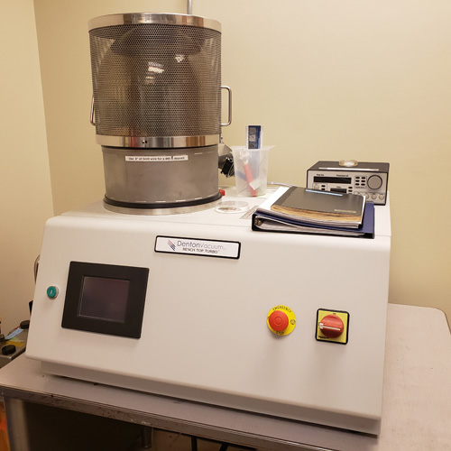
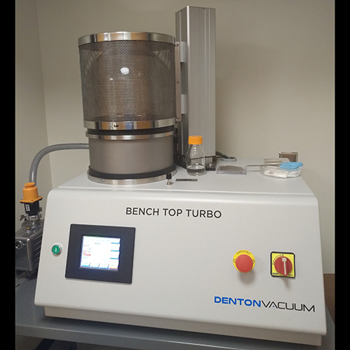
Denton Benchtop Turbo Carbon/Gold Evaporator
Cherry Logan Emerson Hall. Suite E114
- This instrument is used to apply thin layers of carbon or gold on surfaces and to prepare samples for SEM imaging.
- An Inficon SQM-160 rate/thickness monitor allows the application of thin and ultrathin layers of carbon or gold to specimen support grids or other support materials.
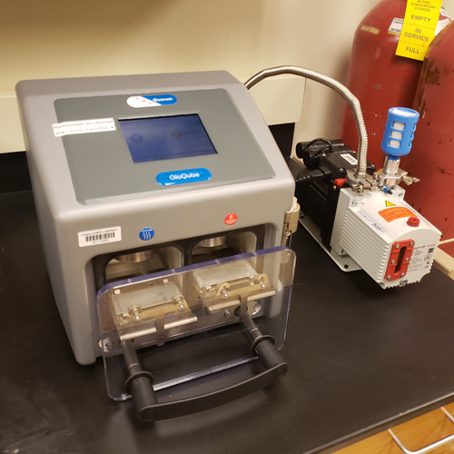
GloQube Glow Discharger
Cherry Logan Emerson Hall. Suite E114
- This instrument is used for cleaning and glow discharging TEM grids and modifying surfaces.
- Various programs are available to generate specific conditions on the grid surface for various TEM applications.
- Two-chamber system for separated in-air and in-vapor processing, without cross-contamination between chambers.
- The second isolated chamber can be used to create more specialize conditions. With automatic vapor delivery, controlled amounts of chemical vapors can be incorporated in the plasma.
Imaging Data Processing and Analysis
Cherry Logan Emerson Hall. Suite E109
- An iMac workstation is available for users to carry out image processing.
- Through a direct connection with an SBGrid-supported, TensorEX, 4 GPU, Linux workstation, users can access structural biology processing software including single particle, tomography, microED, and x-ray crystallography applications.
IEMC Instrumentation - Biochemistry Connector Site - Microscopes
ThermoFisher Talos Arctica 200 kv TEM
Biochemistry Connector. Suite G239
- The ThermoFisher Talos Arctica Field Emission TEM has a BioQuantum Gatan K3 direct electron detector.
- Images are acquired using a Gatan Image Filter system (GIF) which is especially useful for high-resolution cryo-electron tomography, cryo-electron microscopy of thick samples, and to improve contrast on cryo-electron microscopy specimens.
- This instrument is equipped with an autoloader system, capable of loading 12 samples at a time, which can be maintained under liquid nitrogen temperatures for easy sample exchanges.
- Single particle data and tilt series acquisition are done using ThermoFisher EPU and Tomography software and the Serial EM software.
Please contact IEMC (iemc@emory.edu) to request training or use the link below to book this instrument.
Book this microscope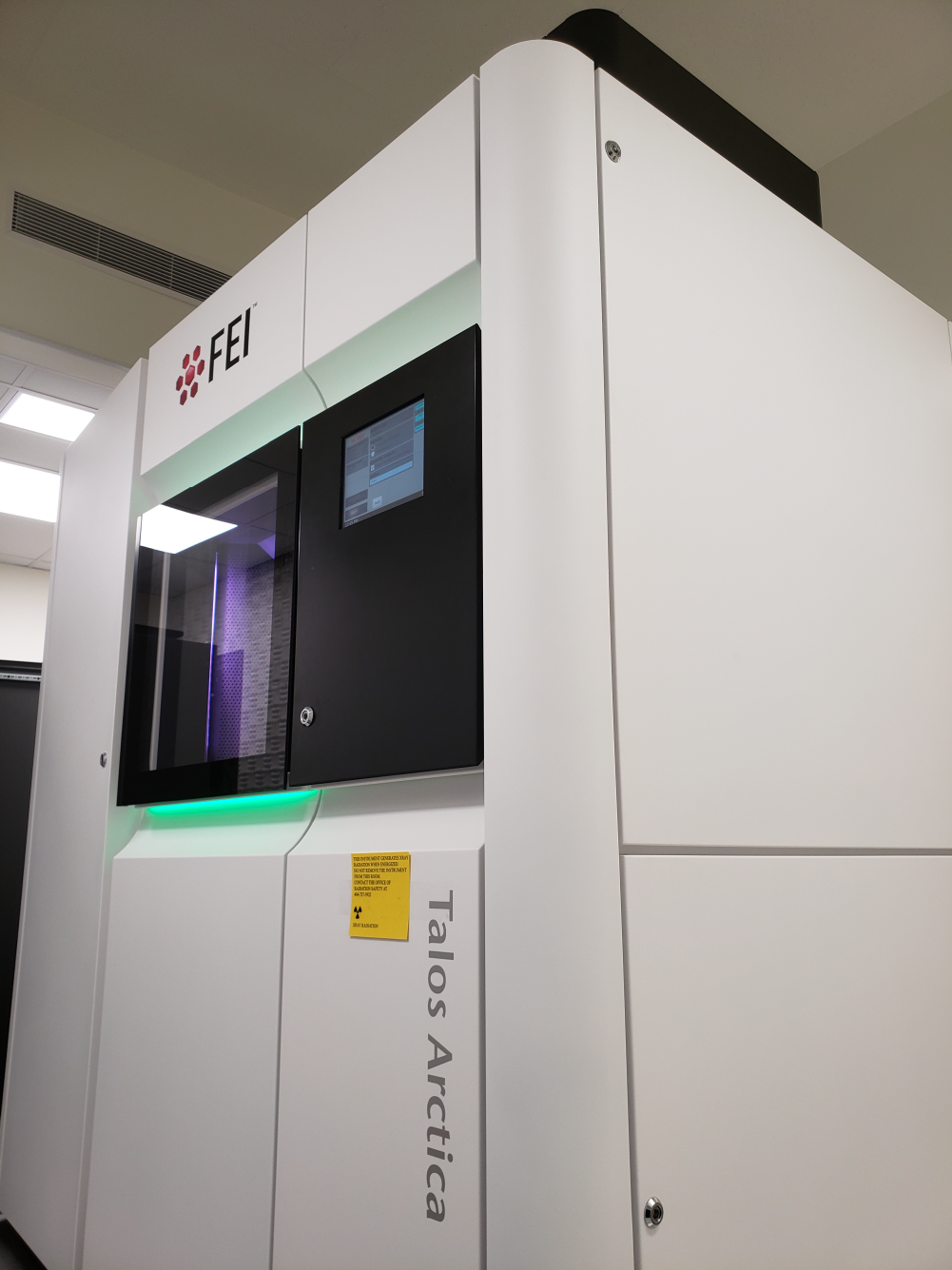
ThermoFisher Talos L120C 120 kv TEM
Biochemistry Connector. Suite G239
- The ThermoFisher Talos L120C, 120 kV, LaB6TEM is equipped with a 4k × 4K Ceta CMOS camera.
- This instrument is capable of cryo-TEM, and room-temperature TEM.
- There are two Gatan cryo holders available for the acquisition of cryo-TEM data, a Gatan 626 and a Gatan ELSA holder.
- Semi-automated data acquisition using Serial EM is available, and a Low Dose function allows for imaging and screening beam-sensitive samples.
- The beam blocker allows for electron diffraction experiments.
Please contact IEMC (iemc@emory.edu) to request training or use the link below to book this instrument.
Book this microscope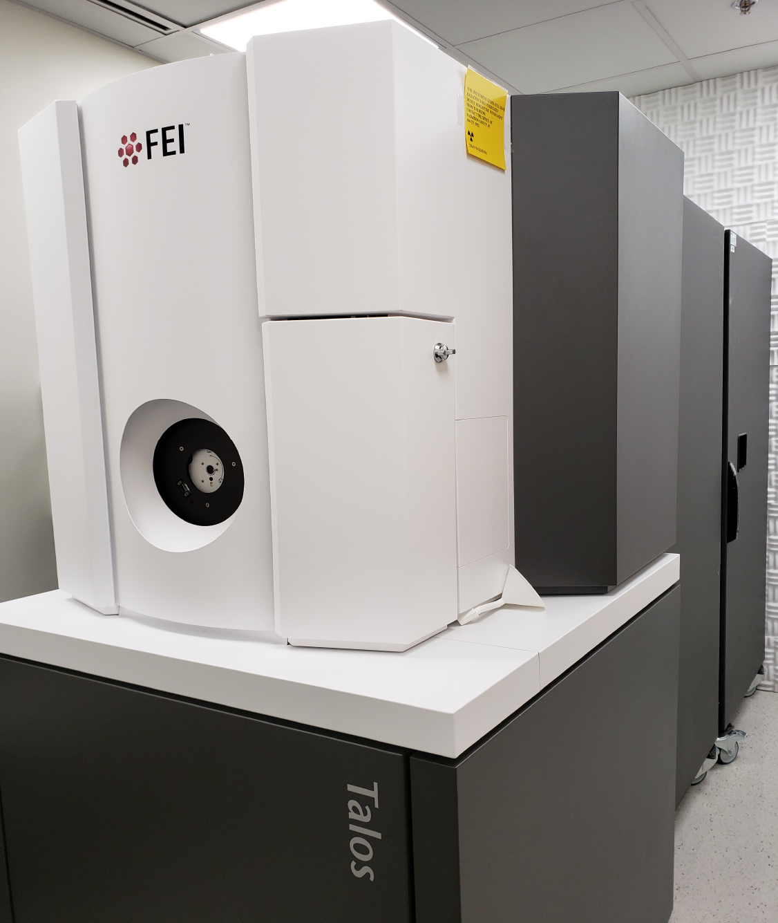
IEMC Instrumentation - Biochemistry Connector Site - Sample Preparation Equipment
ThermoFisher Vitrobot Mark IV
Biochemistry Connector. Suite G236A
- The ThermoFisher Vitrobot Mark IV is used for plunge freezing aqueous solutions, cell suspensions, and on-grid grown cells.
- Vitrification can be done in liquid ethane, liquid propane, or an ethane/propane mix to prepare samples for cryo-TEM, cryo-ET, and microED data acquisition.
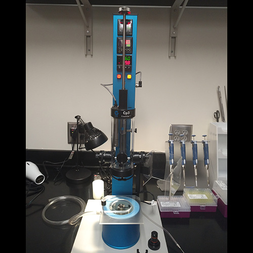
Gatan CP3 Plunger (Model 930)
Biochemistry Connector. Suite G236A
- For plunge freezing aqueous solutions, cell suspensions, and on-grid grown cells.
- Gentle blot for cryo grid preparations.
- Vitrification can be done in liquid ethane, liquid propane, or an ethane/propane mix to prepare samples for cryo-TEM, cryo-ET, and microED data acquisition.
Denton Benchtop Turbo Carbon Evaporator
Biochemistry Connector. Suite G236
- This instrument allows the application of thin layers of carbon on surfaces.
- This instrument can be used to apply carbon on plastic-coated TEM grids which can then be used for negative staining.
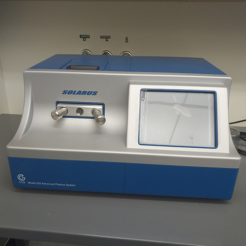
Gatan Solarus Plasma Cleaning System (Model 950)
Biochemistry Connector. Suite G238
- This instrument is used for cleaning and glow discharging TEM grids and TEM cryo and room temperature holders.
- Direct connection to Oxygen, Hydrogen, and Argon sources allows for the combination of these gases to generate surface conditions amenable for a variety of samples for TEM applications.
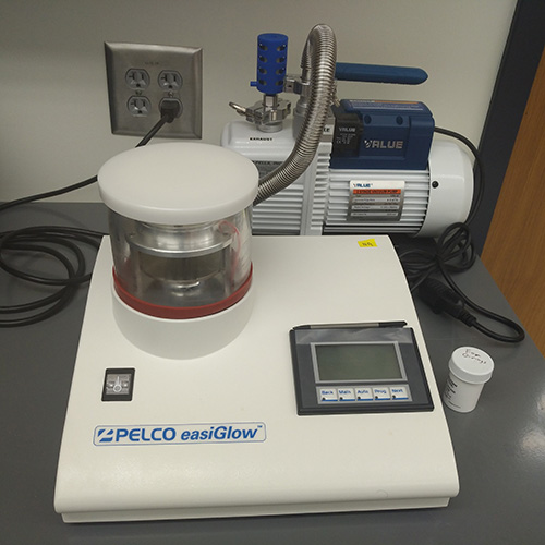
Pelco Easy Glow
Biochemistry Connector. Suite G238
- This instrument is used for cleaning and glow discharging TEM grids.
- Various programs are available to generate specific conditions on the grid surface for various TEM applications.
GloQube Glow Discharger
Biochemistry Connector. Suite G238
- This instrument is used for cleaning and glow discharging TEM grids and modifying surfaces.
- Various programs are available to generate specific conditions on the grid surface for various TEM applications.
- Two-chamber system for separated in-air and in-vapor processing, without cross-contamination between chambers.
- The second isolated chamber can be used to create more specialize conditions. With automatic vapor delivery, controlled amounts of chemical vapors can be incorporated in the plasma.
Imaging Data Processing and Analysis
Biochemistry Connector. Suite G236
- An SBGrid-supported, TensorEX, 4 GPU (NVIDIA RTX A6000, 48GB GDDR6), Linux workstation is available to IEMC users.
- Structural biology processing software includes single particle, tomography, microED, and x-ray crystallography applications.