Emory IEMC Applications and Techniques
Through the Georgia Core Facilities Partnership, IEMC instrumentation is available to researchers from member institutions of the Georgia Research Alliance, which includes Augusta University, Clark Atlanta University, Emory University, Georgia Institute of Technology, Georgia State University, Mercer University, Morehouse School of Medicine, and The University of Georgia.

Single-particle cryo-electron microscopy (SP-cryo-EM) enables high-resolution structural determination of purified proteins, complexes, and nanoparticles without the need for crystallization. By rapidly vitrifying samples and imaging thousands to millions of individual particles, this powerful technique reconstructs 3D structures that reveal molecular architecture, conformational states, and functional mechanisms. At the IEMC, SP-cryo-EM enables researchers to: • Visualize macromolecular complexes in a near-native, hydrated state • Capture multiple conformations and dynamic structural changes • Determine structures of challenging or flexible targets • Accelerate discovery in structural biology, virology, drug design, and nanomedicine From sample optimization and grid preparation to data collection and image processing, our expert team supports the entire workflow—helping transform purified samples into publication-quality structures.
Visit our website at www.cores.emory.edu/iemc www.cores.emory.edu/iemc for more details or contact us at emcore@emory.edu for your electron microscopy needs.
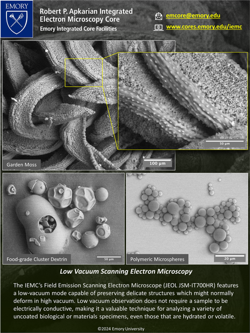
With the low-vacuum mode on our JEOL JSM-IT700HR Field Emission Scanning Electron Microscope (JEOL USA, JEOL Ltd.), we can preserve delicate structures which may normally deform under high vacuum imaging conditions. Using low vacuum we are able to analyze a variety of uncoated biological or material specimens.
Visit our website at www.cores.emory.edu/iemc www.cores.emory.edu/iemc for more details or contact us at emcore@emory.edu for your electron microscopy needs.
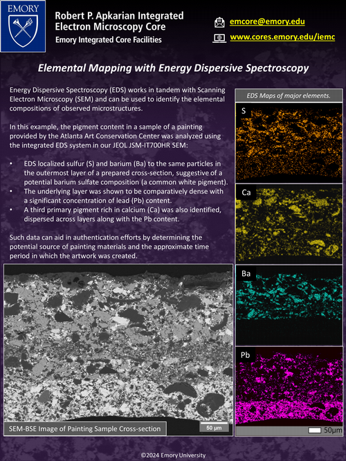
Combining ultrastructural analysis with sample composition has enormous applications in all areas of biological electron microscopy. At the Robert P. Apkarian Integrated Electron Microscopy Core (IEMC), we address this line of research using our JEOL JSM-IT700HR Field Emission Scanning Electron Microscope (FE-SEM), equipped with a JED-2300 Energy Dispersive Spectroscopy (EDS) system (JEOL USA, JEOL Ltd.). This instrument offers advanced capabilities such as image capture, multipoint analysis, line scan, hyperspectral mapping, drift correction, and semi-quantitative analysis. Biological SEM/EDS offers valuable topographic and compositional information of resin/block sectioned or trimmed samples. The examples in this flyer illustrate how SEM/EDS can provide not only ultrastructural information but also data on the distribution of elements in biological samples and their association with ultrastructure.
Visit our website at www.cores.emory.edu/iemc to learn more about our SEM/EDS capabilities or contact us at emcore@emory.edu for your electron microscopy needs.
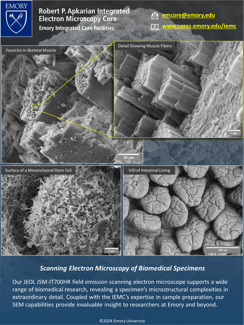
With our JEOL JSM-IT700HR Field Emission Scanning Electron Microscope (JEOL USA, JEOL Ltd.), we analyze biological specimens to uncover their microstructural complexities in detail. The IEMC features high-end sample preparation instrumentation including critical point drying systems and sputter coating equipment, for various applications.
Visit our website at www.cores.emory.edu/iemc or contact us at emcore@emory.edu for your electron microscopy needs.
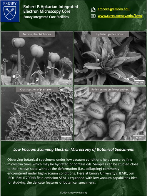
Our JEOL JSM-IT700HR field emission SEM, allows us to investigate botanical specimens under low vacuum conditions, in a near-native state. This capability is enhanced even further by using a low vacuum secondary electron detector which boosts spatial resolution while taking advantage of the low vacuum capabilities to study delicate samples, including plant materials.
Visit our website at www.cores.emory.edu/iemc or contact us at emcore@emory.edu for your electron microscopy needs.
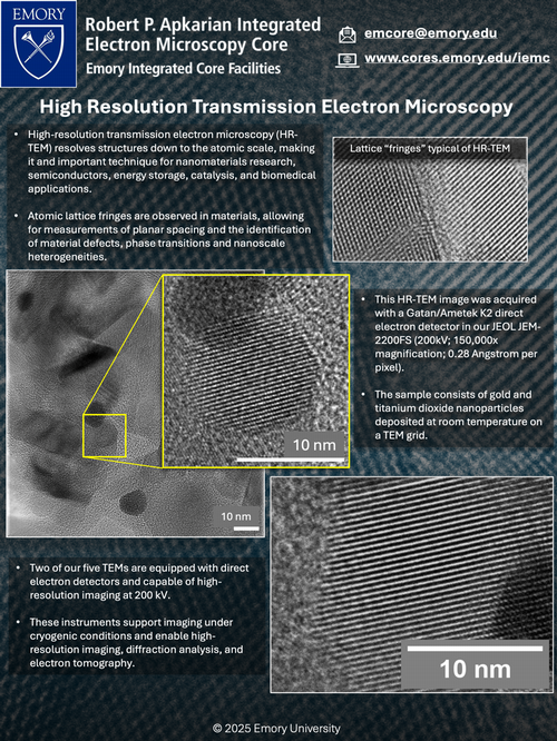
At the Robert P. Apkarian Integrated Electron Microscopy Core (IEMC) at Emory University, we provide state-of-the-art High-Resolution Transmission Electron Microscopy (HR-TEM) capabilities for structural characterization at the atomic scale. Our 200 kV TEMs, equipped with energy filters (Gatan Image Filter, JEOL Omega Filter) and direct electron detectors (Gatan/Ametek K2 and K3, Direct Electron DE20), allow researchers to resolve lattice spacings in inorganic materials. These instruments enable high-resolution imaging, diffraction analysis, and electron tomography, making them essential for nanomaterials research, semiconductors, energy storage, catalysis, and biomedical applications. HR-TEM provides detailed atomic-level characterization, revealing subtle defects, phase transitions, and nanoscale heterogeneities that directly influence material properties and performance.
Visit our website at www.cores.emory.edu/iemc to learn more about our HR-TEM workflows or contact us at emcore@emory.edu for your electron microscopy needs.