Auxiliary devices
Auxiliary Devices
Our standard auxiliary setting for human MRI includes the following items,
| Devices\Site | Purpose | EUH - 3T | HSRB2 - 3T | EP12 - 3T | HSRB2 - 7T |
| Visual Presentation | Present static image, video, and visual paradigm to MR subject | Cambridge Research Systems BOLDScreen 32" LED | Cambridge Research Systems BOLDScreen 32" LED | Cambridge Research Systems BOLDScreen 24" LED | Cambridge Research Systems BOLDScreen 32" LED |
| Intercom | MR technologist/subject communication | Avotec Silent Scan® | Avotec Silent Scan® | Avotec Silent Scan® | Siemens Scanner room roof top speaker |
| Acoustic Presentation | Present audio stimulation | SensimetricsModel S14 Earphones | SensimetricsModel S14 Earphones | N/A | SensimetricsModel S15 Earphones |
| Button Response | Record subject response during fMRI | Current Designs fORP Psychology Software Tools Celeritas® | Psychology Software Tools Celeritas® | Current Designs fORP | Psychology Software Tools Celeritas® |
| Eye tracking | Track eye movement during fMRI and rsfMRI scans | MRC eyetracker, single eye | MRC eyetracking camera | MRC eyetracker, single eye | MRC eyetracker, high speed, both eyes |
| Vital Sign Monitoring | SPO2 | Siemens wireless, BIOPAC | Siemens wireless, BIOPAC | Siemens wireless, BIOPAC | Siemens wireless, BIOPAC |
| EtCO2 | BIOPAC | BIOPAC | BIOPAC | BIOPAC | |
| ECG | Siemens wireless | Siemens wireless | Siemens wireless | Siemens wireless | |
| Respiration | Siemens wireless | Siemens wireless | Siemens wireless | Siemens wireless | |
| EEG | fMRI compatible EEG | Brain Products EEG | N/A | N/A | N/A |
| Infusion Pump | Contrast agent injection | IRadimed | IRadimed | IRadimed | N/A |
| GSR | Skin resistance | BIOPAC | BIOPAC | N/A | BIOPAC |
| EMG | Electromyography | N/A | BIOPAC | N/A | BIOPAC |
| Voice Recorder | Voice response | Avotec | Avotec + Optical microphone | Avotec | N/A |
| Inhale administration | Administration of premixed air with alternative O2/CO2, and other contents | Airgas + homemade tubing/mask | Airgas + homemade tubing/mask | Airgas + homemade tubing/mask | Airgas + homemade tubing/mask |
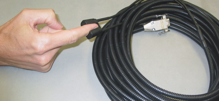
The CORE is open to user provided MRI compatible auxiliary devices, like in-bore exercise setting, force and torque sensors, temperature sensor/gauge, MRI compatible brain stimulator, electric shock electrodes, buzzer, goggle, mouse/joystick, etc.
- SpO2 - The blood oxygen saturation monitor. This appears like a rubber ring attached on one of the index or middle finger. We use this to obtain the heart cycle and blood oxygen saturation level during a scan. These data are usually used to filter out physiological noises in functional MRI data, and find out if the participant is stressed during MR scans.
- Breath cycle. This appears like a belt wrapped around the chest. We use this to obtain the respiration cycle data. These data are usually used to gate the respiration induced organ movement during abdomen scans, and helps to reduce physiological noises during functional scans.
- ECG - electrocardiography. The MR technologist will attach 3-4 receive only self-adhesive electrodes on the participant's chest. These data are usually used to do heart cycle gating for cardiac scans.
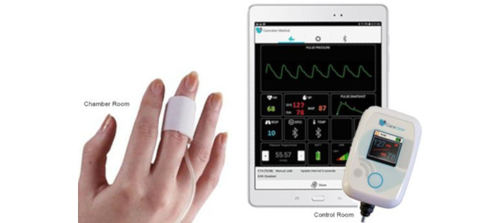
- Blood pressure. This appears like a small plastic piece wrapped on the thumb using Velcro. We use this to continuously monitor the blood pressure. These data are usually used as a reference of the anxiety variation during a scan.
- GSR/EDA - galvanic skin response, electro-dermal activity. This appears like a pair of passive electrodes affixed on the fingers, toes, or other skin location on your arm or leg. It measures the tissue resistance between two electrodes (active, GSR) by introducing a weak electric current one cannot feel, or the voltage difference between the two electrodes (passive, EDA). We use this to evaluate the emotional arousal.
- EtCO2 - the amount of carbon dioxide (CO2) in exhaled air. To do this, a small tubing will be introduced to a location close to the nose of the participant, or a respirator will be mounted on the face. It continuously pumps a small amount of exhaled air into a CO2 percentage sensor. Frequently this measurement is done with breath-able gas administration - a formulated air flow with alternated oxygen, carbon dioxide, and other content, will be introduced through the respirator. We use this information to evaluate the brain function difference when there are different levels of oxygen, carbon dioxide, etc. in the blood stream.
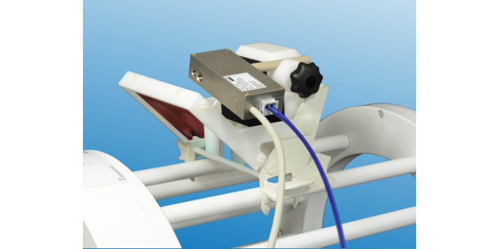
- Eye tracker. To do this, one or two miniature cameras are mounted on a frame over the head coil. It records video of the eye movement and a computer software analyze the video to find out where the participant is looking at on the screen. We use this to find out what the participant is looking at and coordinate it to the brain activities recorded in functional MRI scans.
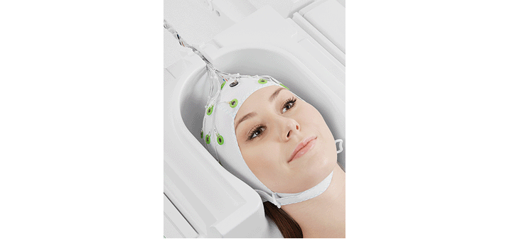
- EEG - electroencephalography. To do this, a technician will tie a hat with 65 electrodes over the participant's head, and apply conductive gels through holes of each electrodes to establish conductivity to the different location on the scalp. This device will record the electric potential through 64 channels from 64 locations on the head. This provides an alternative mapping of brain activities.
- In some projects the participant's voice responses are recorded.
- In some projects the vital signs like heartbeat, respiration, and blood pressure are monitors using a hospital grade vital monitor.