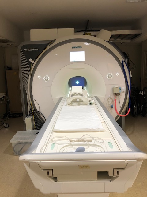
Siemens PrismaFIT 3T at Emory University Hospital
Siemens PrismaFIT 3T at Emory University Hospital
MRI scanners
The Magnetom PrismaFIT whole-body MR system is equipped with a state-of-the art gradient system with
- a maximum (per axis) strength of 80 mT/m and slew rate of 200 T/m/sec
- 64 independent RF receiver channels capable of 204 receiver connections
- a 2-channel RF transmitter.
Multiple MR coils are available, including
- a 64-channel head/neck coil with 52 channels for imaging of the head region
- a 32-channel head-only coil
- a 20-channel head/neck coil with 16 channels for head
- spine array coil
- flexible chest coil
- large and small flexible coil for extremity imaging
- Tx/Rx CP Head Coil for large no-cap head space
- and a 31P dual-tune flexible coil for phosphor spectroscopy
It runs the Siemens Syngo VE11C software. In addition, the scanners are equipped with DirectRF and DirectConnect technology, providing a significant increase in signal-to-noise ratio. The PrismaFIT scanner platform allows efficient acquisition of high-resolution fMRI and DTI images with protocols comparable to those released by the Human Connectome Project. Furthermore, it is equipped with multinuclear spectroscopy and additional shim power for improved magnetic resonance spectroscopy. A number of advanced research sequences are also available, including
- Vessel Size Imaging
- quantitative Arterial Spin Labeling
- Diffusion Spectrum Imaging (for High Angular Resolution Diffusion Imaging)
- Simultaneous Multi-Slice EPI (allowing for sub-second high-resolution whole-brain fMRI data acquisition)
- 4D phase contrast MR for measuring time resolved flow velocity
- displacement encoding (DENSE)
- multi-echo and ultra-short echo time sequences
With our master research agreement with the vendor, advanced work-in-progress MR sequences from the vendor, collaborators from other institutions, or locally developed sequences can be deployed.
Stimulus and response system for functional MR
This MR scanner is equipped with peripheral systems for fMRI. Stimulus/response controls for behavioral tasks concurrent with fMRI are supported by an array of hardware specifically designed to allow investigator flexibility and precision.
- Visual presentation is provided by a high resolution LCD projection system (1400x1050 SXGA, 4200 lumens, 1300:1 contrast ratio) delivered from the back of the suite onto a custom fit screen mounted within the bore behind the participant's head.
- Audio presentation is provided by an Avotec Silent Scan 3100 that has been calibrated to maintain sound pressure levels that are dependent directly on input (flat frequency response +/- 4dB, 200-4500Hz range).
- A fiber-optic ergonomic bilateral button response system, as well as other custom response devices like joysticks, steering wheels, wands from Current Designs are equipped for collecting responses from fMRI subjects.
Other Equipment
Peripheral equipment, including
- computers and software for paradigm generation
- setup for stimulus presentation
- devices for recording behavioral data and physiological parameters including
- heartbeat
- respiration
- blood pressure
- eye movement
- ECG
- EEG
- EMG
Stimulus generation and presentation setup allows us to present acoustic, electric, and vibrational stimuli and oral and venous administration of liquids.
Setup for response via button box, keyboard, mouse, speech, eye movement, and grip force has also been established.
We are also equipped with an electronic shop and a small machine shop, providing the capability to fabricate custom MRI coils, animal holders, and special purpose stimulation devices.