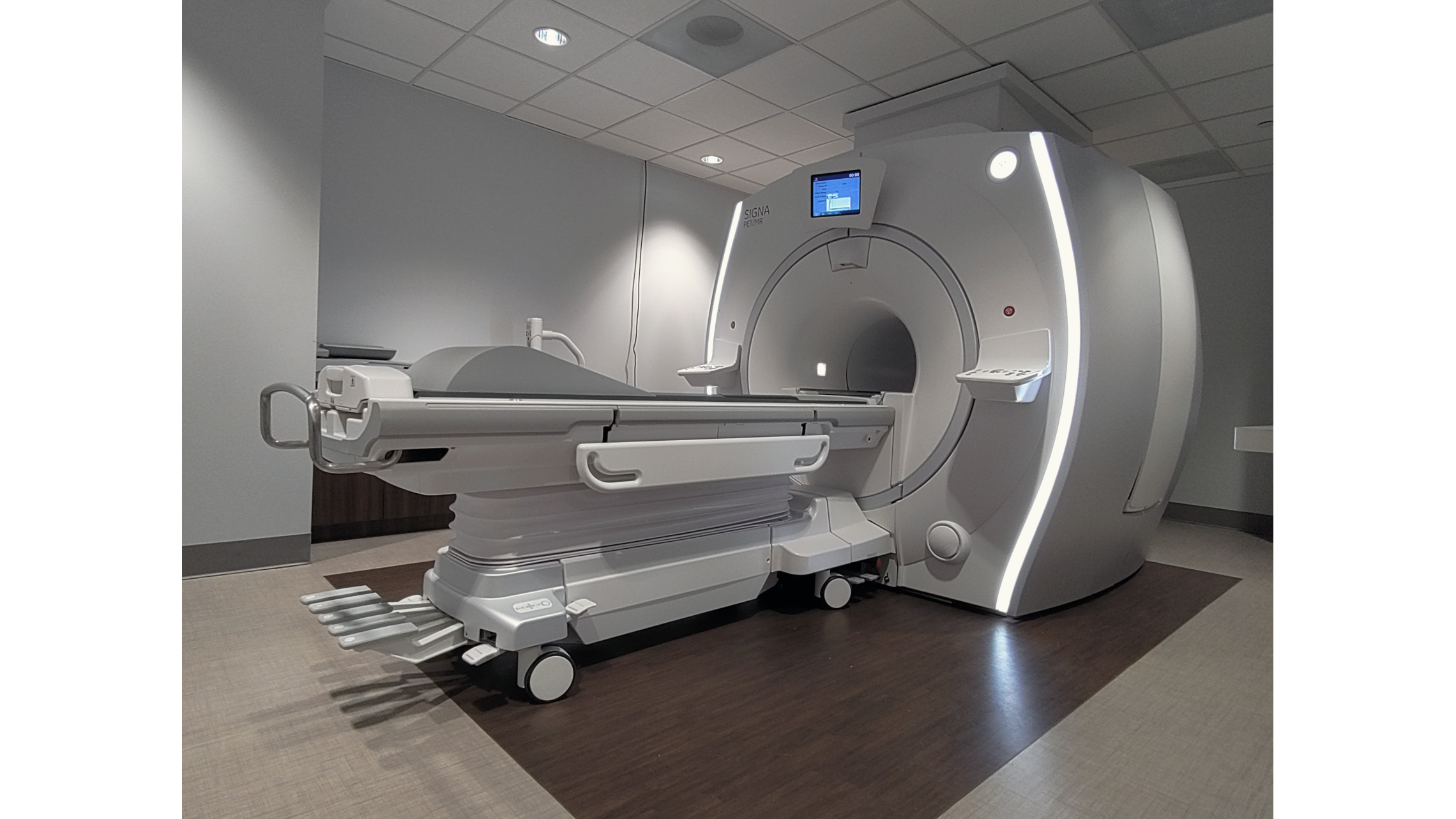
GE SIGNA PET/MRI System
A 3 Tesla PET/MR system from GE Healthcare is shared between Center for Systems Imaging Core (CSIC) and Emory Healthcare to support both research study and clinical scans.
A 3 Tesla PET/MR system from GE Healthcare is located in a translational area within the Emory Clinic (building C - Winship) and supports both research studies and clinical use via a Shared Use Agreement between EHC and CSIC. The system was purchased with funds from an NIH S10 high-end instrumentation (HEI) grant, and supplemented with Emory Healthcare funds. Investigators that need to utilize the dedicated research allotted time on the PET/MR should follow the process to onboard their study with CSIC.
The GE SIGNA PET/MR 3.0T system with Quantworks features a simultaneous time of flight (TOF) PET imaging integrated with whole body 3.0T MRI scanner in a 60 cm bore. The PET system is composed of 45 LBS-Lutetium based scintillator rings (20,160 total crystals) and 28 Silicon Photomultiplier Modules with 21cps/kBq sensitivity. The PET ring is integrated into the MR RF body coil structure. The time resolution is <0.4 ns with a coincidence window of 4.57 ns. Axial field of view is 25.0 cm, the trans-axial field of view is 60 cm. Trans-axial resolution is 3.7-4.2mm and axial resolution is 4.8-7.1mm. The PET processing system contains 896 1.15 GHz GPU cores and two six-core Intel Xeon CPUs. The high PET sensitivity offers the potential for additional dose reduction or, alternatively, faster PET scans for the same dose. The exceptional TOF capability offers higher signal-to-noise images and improves PET attenuation correction with truncation correction, compared to non-TOF reconstruction systems. The system is also equipped with a number of viewing and image processing programs, including zero-TE attenuation correction for the head.
The MR system is a 3.0 Tesla GE SIGNA MR750W equipped with gradients capable of a Peak Amplitude 44 mT/m and a Peak Slew Rate 200 T/m/s and the state-of-the-art MRI capabilities including multi-nuclear spectroscopy. Main field homogeneity is <0.500 ppm over 40 cm field of view (FOV). It has 136 coil input ports that support up to 30 simultaneous imaging coil elements, including a Head and Neck Unit (HNU), an integrated Central Molecular imaging Array (CMA), a PET/MR 8-channel high resolution head array coils. HNU can be combined with the fixed posterior unit to form a 24-element coil array for head and neck imaging. These specialized coils are designed with low 511 keV photon attenuation to preserve PET image quality. The CMA coil is fully-integrated into the patient bore and can be used in conjunction with Upper Anterior Array (UAA) and Lower Anterior Array (LAA) coils for spine and abdomen imaging. The software suite includes packages for advanced neuro, body, oncology and orthopedic, cardiovascular, and pediatric application.
This PET/MR scanner supports PET/MR imaging needs in oncology, neurology, and cardiology with true simultaneous PET and MR acquisition for structural, molecular and functional imaging of brain, spine, body, and musculoskeletal and vascular systems. A robust, automated, MR-based attenuation correction (MRAC) procedure creates attenuation maps to correct the PET data.
The MR system is licensed with GE MAGiC synthetic MRI post-processing package. It generates different contrast weightings the parametric maps of tissue properties obtained from the same acquisition. The MAGiC package is able to synthesize T1 weighted, T2 weighted, IR, FLAIR, STIR, PSIR, FSE-IR, and TIRM images by setting corresponding TR, TE and TI parameters without rescanning. The MR system is also integrated with Acoustic Reduction Technology (ART), which reduces the acoustic noise and improve the patient environment. The detachable patient table can be positioned head first or feet first. The IntelliTouch technology eliminates the need for laser alignment.