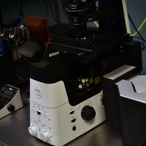
Nikon Crest Optics X-Light V2 L-FOV | inverted confocal | WCI
- Fast and gentle spinning disk confocal
- Large field of view
- Seven laser lines [nm]: 365, 440, 488, 514, 561, 640
- Z-stacks, timelapse, multi-point imaging, tiling
- Live-cell imaging with stage top incubator
- Motorized disk bypass mode for widefield imaging
- Has water immersion objectives (20x/0.95 and 40x/1.15)
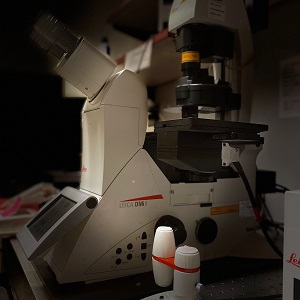
Leica SP8 | inverted confocal | WCI
- Point scanning confocal
- Six laser lines [nm]: 405, 458, 488, 514, 561, 633
- Z-stacks, timelapse, multi-point imaging, tiling
- Live-cell imaging with stage top incubator
- Galvano and 8 kHz resonant scanners for faster and gentler live-cell imaging
- FRAP, FLIP, FRET, photoactivation, reflection
- Inverted microscope body
- Navigator software for tiling
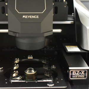
Keyence BZ-X810 | inverted widefield | Winship Cancer Institute
- Automated widefield imaging system
- Four filtercubes: DAPI, FITC, Texas Red, Cy5
- Fluorescence, bright field, color bright field, phase
- motorized stage for tiling and z-stacks
- Reads slides, dishes, and plates
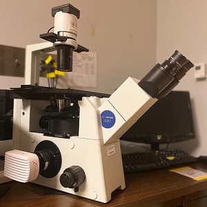
Olympus IX51 | inverted widefield | WCI
- Five filtercubes: DAPI, FITC, TRITC, Texas Red, Cy5
- Fluorescence, bright field, phase
- Five objectives from 2x to 40x
- 1.4 megapixel monochrome CCD
- Inverted
- Use for quick sample checks